Structure of the eye

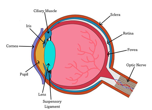
Figure 11.1.a
Source: Sruthi Sridhar
Light can enter the eyes either directly from a source (sun, light bulb) or indirectly through the reflection of an object (also reflected multiple times from various objects). In order to have a clear vision, a single point of light must travel through the structures present in the eye and land on the retina. In this process, refraction occurs as light bends when it passes through varying densities of matter. For example, a drinking straw appears broken on the water’s surface. The structures in the eye are critical for gathering and focusing light so that humans can see clearly.
See detailed images of the eye:
• https://www.aao.org/eye-health/anatomy/parts-of-eye
The cornea is a transparent membrane that covers the eye's surface. The cornea shields the eye and focuses most of the light entering the eye. The cornea has a fixed curvature equivalent to if a camera lacked the ability to adjust focus.
The aqueous humour, a clear, watery fluid, is the next visible layer. This fluid is replenished regularly and provides nutrients to the eye. The light from the visual image then enters the eye's interior through a hole called the pupil, which is located in the iris. Iris is a circular muscle which is the colored part of the eye. The iris can adjust the size of the pupil, allowing more or less light into the eye, which helps focus the image. People try to achieve the same thing by squinting.
Muscles behind the iris suspend a clear structure called the lens. The cornea begins the focusing process, which is completed by the flexible lens. The lens changes its form from thick to thin during visual accommodation, allowing it to concentrate on close or far away things. The lens' thickness variation helps it to project a sharp image onto the retina.
After passing through the lens, light travels through a large, open region called the vitreous humour, which is filled with a clear, jelly-like fluid. This liquid, like the aqueous humour, nourishes and shapes the eye.
The retina, a light-sensitive area at the back of the eye, has three layers: ganglion cells, bipolar cells, and rods and cones, which are unique receptor cells. These photoreceptors respond to various wavelengths of light. The retina is the final stop for the light within the eye.
While the retina absorbs and processes light, the rods and cones are responsible for receiving photons of light and converting them into neural activity for the brain. The signal is then transferred to the bipolar cell (a form of interneuron) and then to the retinal ganglion cells, whose axons form the optic nerve.
Different aspects of vision are controlled by the rods and cones. Each eye has 6 million cones, with 50,000 of them having a private line to the optic nerve (one bipolar cell for each cone). This means that the cones are the visual acuity receptors, or they have the capacity to discern fine details.
Rods (approximately 100 million in each eye) are located across the retina except in the fovea, where they are concentrated. Rods are sensitive to brightness changes but not to a wide range of wavelengths. Thus, they can only see in black and white and shades of grey. As multiple rods are linked to a single bipolar cell, the brain interprets a photon of light to stimulate an entire area of rods. The visual acuity (sharpness) is low because the brain does not know which region (which particular rod) is transmitting the message. As a result, things appear fuzzy and greyish in low-light situations like twilight or dimly lit rooms. Rods are also responsible for peripheral vision because they are located on the retina's periphery.
Cones can be found throughout the retina, although they are more abundant in the centre, where there are no rods (the area called the fovea). Cones also require a lot more light than rods to function; thus, they perform best in bright light, which is also when people see objects the best. Cones are responsible for color vision because they are sensitive to different wavelengths of light. While the eye supports many visual processing activities, such as the perception of lightness and brightness, due to lack of space, here we will deal with one such phenomenon of vision–the perception of color.
Perception of Color
In the 1800s, two hypotheses were proposed concerning how people perceive colors. The trichromatic ("three colors") theory is the first. This concept advocated three sorts of cones: red cones, blue cones, and green cones, one for each of the three primary colors of light. The theory was first proposed by Thomas Young in 1802 and later refined by Hermann von Helmholtz in 1852. According to the trichromatic hypothesis, color shades correspond to various amounts of light received by each of the three types of cones. The message from these cones is subsequently sent to the brain's vision centres. The visible color is determined by the combination of cones and the rate at which they are fired. For example, if the red and green cones fire at high enough rates in response to a stimulus, the person perceives yellow.
The trichromatic hypothesis appears to be more than enough to explain how people perceive color. However, this theory cannot account for an intriguing phenomenon. Let us experiment and focus on the union jack for at least a minute. Right after a minute, look at the white box next to it. What do you see?

Figure 11.1.b
Source: Sruthi Sridhar. Recreated from Ciccarelli and White, 2018 (page 144)
You should have seen an afterimage of the flag. An afterimage is a visual sensation that lasts for a short period after removing the original stimulus. Furthermore, the person would notice that the flag's colors are all wrong in the afterimage: green for red, black for white, and yellow for blue.
The second theory, known as the opponent-process theory, which Ewald Hering proposed in 1874, explains the afterimages phenomenon. The opponent-process theory has four fundamental colours: red, green, blue, and yellow. Each hue is arranged in pairs, with each pair member acting as an opponent. The color red is coupled with the color green, and the color blue is paired with the color yellow. When one pair is overly stimulated, the other is inhibited and unable to function; therefore, there are no reddish-greens or blueish yellows.
So, how might such a pairing result in color after images? Some neurons are excited by light from one part of the visual spectrum and inhibited by light from another part of the spectrum. Let us imagine we have a red-green ganglion cell in the retina. The cell will have a very low baseline activity when exposed to white light. However, if there were a red light, there would be increased cell activity, and we would perceive the color red. If the cell is stimulated by a red light for an extended period, it becomes exhausted. When the red light is replaced with white light, the exhausted cell responds even less than before. Therefore, we see the color green, which is related to this reduced cell response.
The Visual Pathway
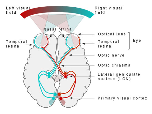
Along with the visual processing occurring in the eye, the visual system consists of the optical pathway and connects the eye to the brain.

Source: Wikimedia
The retina is connected to parts of the brain called the primary visual cortex (see details in the picture). The left and right visual fields can be distinguished when light enters the eye. The left half of each eye's retina receives light from the right visual field. The right half of each retina receives light from the left visual field. Because light passes through the cornea and lens in a straight line, the image projected on the retina is actually upside down and inverted from left to right compared to the visual fields. The right visual cortex receives information from the left visual field and vice versa. The point of crossover is the optic chiasm.
The process of vision involves much beyond the concepts listed here. The above are just a sampling of some workings of the visual system, which have been explored in greater detail in this course. However, vision science remains a major area of research and can serve communication designers' detailed and reflexive understanding of the visual process.

